Take a look at the pictures shown in figures 1.1 through 1.3. They represent what happens when a gross stretch is applied to a muscle. The therapist is holding two resistance bands tied together—one red, the other black. The red resistance band is extremely stretchy; the black is tough and less stretchy. The red resistance band represents normal, healthy muscle tissue; the black resistance band represents an area of tight muscle tissue. Together these bands represent one whole muscle. Look at what happens in figure 1.1 when the therapist moves his right hand. Which part of the muscle does the stretching—the pliable (red) part or the tough (black) part? Clearly, the pliable band is doing the most stretching.

Figure 1.1 Notice which band is doing the stretching.
Now look at figure 1.2. What happens when the therapist moves his left hand? Which part of the muscle stretches the most—the pliable (red) part or the tough (black) part? Again, the pliable band is doing most of the stretching.
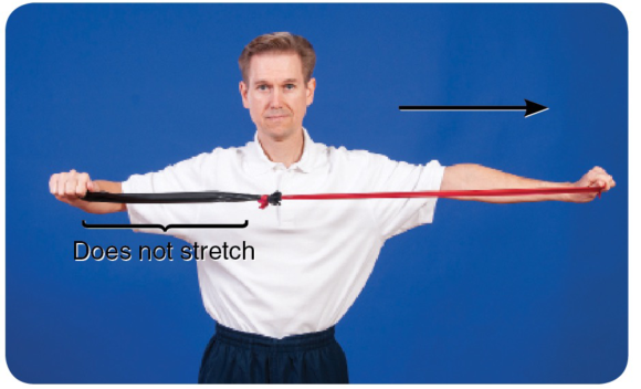
Figure 1.2 Which band is doing the stretching now?
Finally, notice what happens when the therapist moves both his right and left hands apart so they are equidistant (figure 1.3).
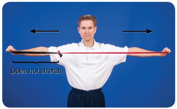
Figure 1.3 Even with an equidistant stretch, the more pliable band does the most stretching.
You can see from the illustrations that the pliable part of the muscle (the red band) does most of the stretching, irrespective of which end of the muscle is moved. To target the less pliable part of the muscle—the area of palpable tightness—you need to localize the stretch. This is exactly what STR does.
To localize the stretch, you need to ‘fix’ part of the muscle against underlying structures to create a false insertion point. The fixing—described throughout this book as a lock—prevents some parts of the muscle from moving and is achieved when a therapist uses his or her own upper body or a massage tool. When a muscle is stretched, its insertion points are moved apart from one another; that is, the area of tissue between the insertion points stretches. Creating false insertion points results in a more intense stretch in some parts of the muscle.
Look at figure 1.4a, which is an illustration of the soleus. You probably already know that the soleus originates from the posterior shaft of the tibia and inserts into the calcaneus. When resting in the prone position, the foot naturally falls into plantar flexion (figure 1.4b). If you pull up your toes (dorsiflexing your foot and ankle), it stretches the muscles of the calf (which are the plantar flexors). Dorsiflexion is therefore a way of applying a gross stretch to the soleus and may be achieved passively, as illustrated in figure 1.4c.
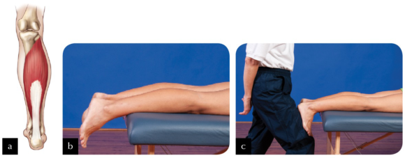
Figure 1.4 (a) The soleus; (b) the ankle falls into plantar flexion in the prone position; (c) performing a passive calf stretch.
Now look at figure 1.5a. Imagine locking the muscle to the tibia slightly distal to its actual origin (lock A; figure 1.5b). Can you see that if you were to stretch the muscle now (figure 1.5c), only those fibres running from the new origin (lock A) to the calcaneus would be able to stretch? Would you agree that, providing you are able to dorsiflex through the same range of motion as in the first stretch, greater force has been placed on those fibres being stretched? This occurs because the small amount of muscle tissue superior to lock A is no longer being stretched.

Figure 1.5 (a) Locking the soleus muscle slightly distal to its actual origin (lock A); (b) applying the lock; (c) performing the stretch.
Now look at figure 1.6a. A second imaginary origin (lock B) for the soleus is even more distal on the tibia, broadly locking it to the underlying structures (figure 1.6b). Performing a stretch now (figure 1.6c) will place even greater tension on the stretching fibres than if the lock had remained at lock A.
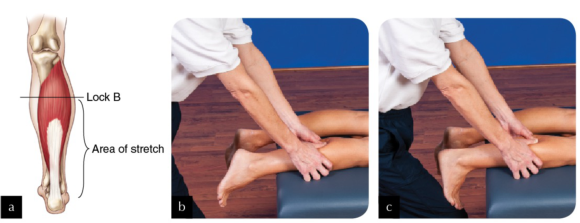
Figure 1.6 (a) Locking the soleus more distal on the tibia (lock B); (b) applying the lock; (c) performing the stretch.
Finally, you could create a third false origin (lock C) yet more distal to the actual origin (see figures 1.7a and 1.7b). In this example, only the most distal portion of the soleus stretches when the foot and ankle are dorsiflexed (figure 1.7c).

Figure 1.7 (a) Locking the soleus even more distal on the tibia (lock C); (b) applying the lock; (c) performing the stretch.
In reality it is not possible—or advisable—to lock the entire breadth of the muscle, but this is the principle behind how STR works. An alternative is to apply a specific rather than a broad lock—for example, on the biceps brachii, as illustrated in figure 1.8. The areas of muscle fibre distal to each of the locks are put under greater stretch each time the elbow is extended. To understand this concept of a specific stretch, think of muscle fibres as the strings of a guitar. Placing your finger across all of the strings, as in the previous example of the soleus, is quite different from placing your finger across one string, as in the case of using your elbow to apply a lock to the biceps. For a start, it is quite difficult to exert the same pressure across all strings that you would use to fix just one string. When playing the guitar, if you use the tip and pad of your finger to fix just one string, with one specific lock, only that string is affected, yet it is affected intensely. However, if you use more of your finger in an attempt to make a lock across all of the strings, you affect all of the strings when you play, though perhaps not as intensely.
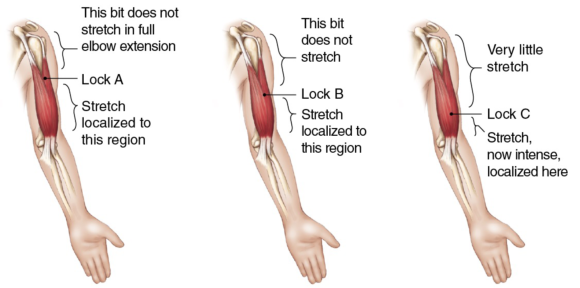
Figure 1.8 In applying specific locks, the areas of muscle fibre distal to each of the locks are put under greater stretch each time the elbow is extended.