Left atrial enlargement (LAE), right atrial enlargement (RAE), and biatrial enlargement can be observed on the 12-lead resting ECG and are characterized by changes in P-wave duration and morphology. Left ventricular enlargement (LVH), right ventricular enlargement (RVH), and biventricular enlargement can be observed on the resting 12-lead ECG and are characterized by changes in the QRS complex, ST segment, and T-wave duration and morphology. Often, ventricular enlargement is accompanied by atrial enlargement with characteristic features on the ECG. This section examines each of these conditions and provides supporting ECGs to illustrate their presentations.
Atrial Enlargement
Dilatation of the chamber or hypertrophy
of the cardiac muscle surrounding the chamber results in characteristic changes on the ECG. Dilatation and hypertrophy often occur simultaneously and are commonly caused by pressure or volume overload. Exceptions to this are the heart muscle diseases including hypertrophic cardiomyopathy (HCM) and dilated cardiomyopathy (DCM).
Right Atrial Enlargement (RAE)
RAE causes an increase in the electrical activity resulting in a greater P-wave voltage (amplitude) and a shift of the P-wave axis to a more rightward position, often toward a vertical axis. In RAE the P wave is abnormally tall (>0.25 mV) with a normal duration (
Following are common causes of RAE:
- Acute pulmonary disease: Bronchial asthma and pulmonary embolism
- Chronic pulmonary disease: Emphysema and bronchitis
- Congenital heart disease: Pulmonary valve stenosis, atrial septal defects, and tetralogy of Fallot
Left Atrial Enlargement
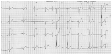
LAE increases the time required for the action potential to spread across the atria resulting in an abnormally wide (>0.12 s) P wave that has either a normal or increased amplitude. The delayed depolarization can result in a humped or notched P wave commonly termed P mitral (this term technically refers only to a humped or notched P wave that is the result of mitral valve stenosis). Occasionally, a biphasic P wave is observed in lead V1 with an initial positive deflection and a wide (>0.04 s), deep (>0.1 mV) terminal portion (see figure 4.31).
Following are common causes of LAE:
- Congestive heart failure associated with all causes including ischemic heart disease and cardiomyopathy
- Valvular heart disease: Aortic stenosis, aortic regurgitation, and mitral stenosis
- Hypertensive heart disease