We start our discussion of dance anatomy and kinesiology in this book by looking at the skeletal system. The skeletal system provides the structural framework of the human body, and its joints permit the varied movements we explore in dance vocabulary. In movements such as the high kick shown in the photo on page 1, bones function in both their support and movement functions. The bones and associated joints of the gesture leg allow for the large movement occurring at the right hip, while those of the support leg are key for providing stability so that the dancer can remain upright despite a very small base of support. The support function of bones requires that they be strong, and understanding of bone remodeling is key for preventing loss in bone strength commonly seen in female dancers. The role of bones in joints is key for understanding and describing human movement. Topics covered in this chapter will include the following:
- Primary tissues of the body
- Bone composition and structure
- Bone development and growth
- The human skeleton
- Joint architecture
- Body orientation terminology
- Joint movement terminology
- Skeletal considerations in whole body movement
The concepts and terminology provided in this chapter will be utilized and applied in more depth in later chapters. Hence, this chapter can serve both as an introduction and as a reference for when this information is readdressed.
The body is composed of four different primary tissues, each with its own particular structure to help it carry out its required functions. These four primary tissues include muscle, nervous, epithelial, and connective tissues. Muscle tissue is characterized by its ability to contract and is found in the heart, in various organs (e.g., in the smooth muscle in the gastrointestinal tract), and in the many skeletal muscles of the body. Nervous tissue is composed of cells (neurons) that are able to generate and conduct electrical messages, as well as other cells (neuroglia) that help support these neurons. Epithelial tissue is composed of cells that fit closely together to form continuous sheets, or membranes, that cover and line surfaces of the body or form glands. Connective tissues generally function to bind, support, insulate, and protect structures and can be further divided into connective tissue proper, cartilage, bone, and blood.
While the first three types of tissues are composed mainly of cells, connective tissue is characterized by the presence of large quantities of nonliving material in the space between connective tissue cells (extracellular matrix; L. extra, outside of), which contains different fibers and other constituents that dictate its form and function. For example, bone has calcium salts within its extracellular matrix that provide it with the type of strength needed to support body weight. Some types of connective tissue proper have closely packed bundles of protein fibers (collagen), giving them the type of strength necessary for their function of binding bone to bone (ligaments) or muscles to bones (tendons). Blood, the atypical connective tissue, has plasma as its extracellular matrix; its fibers become apparent only during the process of blood clotting.
These primary tissues of the body can be grouped together into anatomical or functional units called organs. An organ is a structure that performs a specific function for the body and is composed of two to four of the primary tissues. Examples of organs are the heart and brain. Furthermore, organs that work closely together for a common purpose can be grouped according to a common function into systems, including the skeletal system, muscular system, and nervous system. The skeletal system will be addressed in this chapter, and the muscular system will be addressed in chapter 2. The skeletal system is composed of all of the bones of the body, related cartilages and ligaments, and the joints that connect these bones together.
In the average individual, bone makes up about 15% to 20% of total body weight (Huwyler, 1999). Bone is characterized by its strength and rigidity, and it is one of the strongest connective tissues in the body. Unlike that of other tissues, the extracellular matrix of bone contains calcium salts. These minerals compose about 60% to 70% of bone weight (Hall, 1999; Rasch, 1989) and impart to bone its great compressive (L. pressus, to press together) strength, the ability to resist a force that would tend to push together or crush a bone. This extracellular matrix also contains collagen fibers (G. koila, glue + gen, producing). Collagen imbues bone with its great tensile strength (the ability to resist a pulling force that would tend to pull a bone apart; L. tensio, to stretch) and flexibility. The composition of bone can be compared to that of reinforced concrete, with the collagen playing the role of the steel and the calcium crystals serving the role of the sand and rock. The compression strength of bone is actually greater than that of reinforced concrete (Guyton, 1976), and the tensile strength of very dense bone is estimated to be 230 times greater than that of muscle of a similar cross section (Rasch and Burke, 1978).
The composition of bone allows it to serve in the following key functions.
• Support: Bones provide an internal framework for the body that is essential for stability and form.
• Protection: Some bones protect fragile structures within. For example, the skull helps protect the brain; the rib cage, the heart and lungs; and the pelvic girdle, vital internal organs.
• Movement: Many bones serve as levers to enhance movement capabilities (see Muscles, Levers, and Rotary Motion in chapter 2 [p. 44] for more information). Having long levers in our body allows our limbs to move through a large distance, at a fast speed, or both.
• Blood cell production: Some bones contain tissue (red bone marrow) that is responsible for the production of red blood cells. Red blood cells are vital for the transport of oxygen and carbon dioxide.
• Mineral storage: Various important minerals such as calcium, phosphorus, and magnesium are stored within the bones. When necessary, hormones can stimulate release of some of these minerals into the blood for the body to use. These minerals are vital for important processes such as blood clotting, nerve transmission, muscle contraction, and energy metabolism.
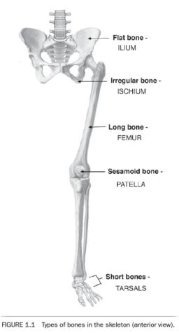
Bones come in a large variety of shapes and sizes. They can be classified according to their shape into the five types described next and illustrated in figure 1.1.
• Long bones are tubular in shape and much longer than they are wide. They are found in the limbs, where they serve as levers to enhance movement. For example, the "thigh" bone, or femur, is a long bone (figure 1.1). Other examples include the clavicles, humerus, radius, ulna, and metacarpals and phalanges of the upper limb or extremity and the tibia, fibula, and metatarsals and phalanges of the lower limb or extremity (figure 1.4). The long bones in the lower extremity are generally larger and stronger to meet their weight-bearing needs, while those in the upper extremity are generally smaller and lighter to meet their role in reaching and in manipulation of objects.
• Short bones are cubical in shape and are found in the upper portion of the hand (carpals; see figure 1.4) and feet (tarsals; see figures 1.1 and 1.4). These bones aid with shock absorption, transmission of forces, and small complex movements.
• Flat bones are relatively thin and flat, but often slightly curved in shape. These bones commonly protect important soft underlying structures (such as the brain), and their shape also allows for extensive attachment of muscles. Examples include the upper portion of the pelvis (ilium) seen in figure 1.1 and the ribs, sternum, scapulae, and some of the bones of the skull shown in figure 1.4.
• Irregular bones do not fall into the preceding three classifications and exhibit complex and varied shapes. Their shape is adapted to special purposes; and they serve a variety of functions including protecting the spinal cord, supporting body weight, transmitting loads, providing sites for muscle attachment, and facilitating movement. Examples include the vertebrae and lower portions of the pelvis (ischium and pubis) shown in figures 1.1 and 1.4.
• Sesamoid bones (G. sesamoeides, like sesame) are bones that form within a tendon. They help protect the tendon from excessive wear due to rubbing against the underlying bone, and they change the angle of the tendon so that the muscle can produce more effective force. Examples include the "kneecap," or patella (figure 1.1), which is encased in the tendon of the quadriceps femoris, and two small bones within the tendon of the flexor hallucis brevis, located under the base of the big toe and discussed in chapter 6. Because these sesamoid bones are relatively flat, many texts include them in the class of "flat bones" just described, while other texts put them in a class of their own.
Bone does not have uniform composition. For example, the relative percentage of mineralization varies between bones, as well as within a given bone to help it better serve its functions. In general, bones have an outer layer of very dense bone called compact bone and an inner layer of less dense bone called spongy, trabecular, or cancellous bone. The compact bone provides strength and stiffness. Cancellous bone (L. a grating, lattice) contains many open spaces between thin processes of bone (trabeculae). These trabeculae (L. trabs, a beam) form a type of latticework that corresponds to the lines of stress occurring within the bone. This architecture provides bones with additional strength and shock-absorbing capacity, while allowing the bones to be much lighter than if they were composed solely of compact bone.
The compact bone, cancellous bone, and other structures present in a typical long bone are shown in figure 1.2. Learning these structures is key for understanding bone growth and health. The shaftlike part, called the diaphysis (G. a growing between), has thick walls of compact bone and a hollow cavity called the medullary cavity (L. marrow). The layer of compact bone thins toward the extremities of the long bone. The enlarged ends of the bones are called the epiphyses (G. epi, upon + physis, growth). These broadened epiphyses provide extensive area for muscle attachment. They also offer larger surface areas for articulation with adjacent bones, enhancing joint stability. The surfaces of the epiphyses that actually come in contact with opposing bones are covered with a thin layer of specialized connective tissue called articular cartilage. Articular cartilage helps lessen forces and allows joints to move more smoothly (see Synovial Joints on pp. 12 and 13 for more information). Rather than housing a hollow cavity, the epiphyses are filled with cancellous bone. The spaces of both the cancellous bone and the medullary cavity are filled with a soft, fatty substance called bone marrow. Some of this marrow (red marrow) is the type that is vital for making red blood cells.
In bone that is still growing, there is a plate of cartilage separating each epiphysis from the diaphysis.
This is an excerpt from Dance Anantomy and Kinesiology.