Muscle damage can result from contusions, ischemia, lacerations, mechanical strain, drug toxicity, and idiopathic causes, as in fibromyalgia or compartment syndromes. Iatrogenic muscle damage (secondary to surgical procedures) is commonly encountered in physical therapy, as it is often necessary for surgeons to dissect muscles and fascia to reach the underlying structures. Treatment is determined by the type of injury and is directed at the suspected underlying problems. The most common causes of muscle injuries are (1) "muscle strains" induced by lengthening contractions and (2) contusions.
Muscle strains are among the most common complaints treated by physicians (Garrett, Jr., 1996). Muscle strains account for the majority of sport-related injuries (Zarins and Ciullo, 1983; Brockett et al., 2001), as well as a significant proportion of low back pain (Bartleson, 2001; Glass, 1979). Therefore, the symptoms associated with these injuries have a significant economic impact on both the individual and society as a whole. When an activated muscle lengthens because the external load exceeds the tension generated by the muscle contraction, its action is termed a lengthening ("eccentric") contraction. Although lengthening contractions require less energy, the force generated during a maximal lengthening contraction is approximately twofold the force developed during a maximal isometric contraction; therefore lengthening contractions are more likely to produce damage than either than isometric or concentric contractions (Hunter and Faulkner, 1997). The basic mechanism of higher force generation during a lengthening contraction is not understood. The difficulty of explaining the enhanced force from a lengthening contraction lies in the fact that the force produced is greater than the sum of the measured active force (from an isometric contraction) and the passive force at that given muscle length.
Lengthening contractions are physiologically relevant (Cavagna, 1977; LaStayo et al., 2003) and often occur without causing damage. They produce high forces, which is a goal of strength training (the overload principle). This is evident in strengthening protocols that use lengthening contractions, or "negatives," to increase strength. Not only can lengthening contractions produce more force than other types of contractions, but they can do so at a reduced oxygen requirement (Lindstedt et al., 2001; LaStayo et al., 1999). Thus, the application of exercises using lengthening contractions in elderly people is appealing, as high metabolic demand is sometimes not wanted in this population. Although greater strength gains are achieved using lengthening contractions in training, many studies have shown that the high force produced by eccentric muscle contractions can cause subsequent pain and damage (Armstrong et al., 1983; Brooks et al., 1995; Friden and Lieber, 2001; MacIntyre et al., 1996).
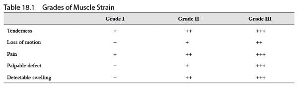
For clinical purposes, a muscle strain can be categorized into three different levels of severity, or grades (table 18.1).
• A grade I strain is a mild strain that is symptomatic (tenderness and mild pain) but results in no impairment (full range of motion and no loss of strength).
• A grade II strain results in moderate impairment, marked tenderness, decreased range of motion of the associated joint, and a noticeable loss of strength.
• A grade III muscle strain results in immediate pain, as well as a palpable defect in the muscle surrounded by swelling (edema), indicating rupture of fibers or the whole muscle.
Although the underlying mechanisms of the different grades of muscle strains may be different, the grades are often reported to represent "the amount of torn fibers." Acute muscle strains can be detected with computed tomography or magnetic resonance imaging (MRI) methods (Speer et al., 1993), but diagnosis is typically made based on physical exam and patient history.
It is important to identify symptoms derived from structures above and below the injury. For example, a condition such as sciatica that causes referred pain down the posterior thigh and leg could be misdiagnosed as a chronic grade I hamstring or calf strain. Tenderness of the muscle-tendon unit-not typically present with referred pain-is a hallmark of muscle injury, and the amount of tenderness increases with the grade of strain. Increased pain in the muscle with isometric resistance applied to oppose the muscle action is another indication of injury.
Muscle injuries are typically self-limiting and do not require imaging, but as this noninvasive technology continues to improve and imaging (e.g., MRI) becomes more commonplace, it may play a role in rehabilitation planning and prognosis (Blankenbaker and De Smet, 2004). Plain films, or X rays, are not very useful for imaging muscle pathology unless heterotopic bone formation has occurred within the muscle (see later discussion of myositis ossificans). Unlike X rays, MRI has high sensitivity to the hemorrhage and edema that follow muscle injuries. This, together with the capability to evaluate multiple anatomic planes, makes it the most suitable technique to evaluate muscle injuries. Muscle strains are revealed best by T2-weighted images, which optimize contrast between injured muscles with edema (increased signal intensity) and normal uninjured muscles (the T1 and T2 relaxation times define the way the protons in tissues revert back to their resting states after an initial radiofrequency pulse) (see figure 18.3). Imaging of muscle injuries does not, however, provide information regarding the underlying mechanisms of injury or the cellular processes that are taking place.